
The nervous system begins as a midline thickening of ectoderm dorsal to the notochord to form a neural plate. The neural plate invaginates, then folds into a neural tube in a process known as neurulation. Problems in this process give rise to neural tube defects such as spina bifida.
Nervous system patterning
CNS patterning
Pattering of the neural tube occurs in three dimensions to create the CNS.
Anterior-posterior axis
- Regionalisation: The neural tube becomes regionalised – anteriorly into the forebrain, midbrain & hindbrain and posteriorly into the spinal chord
- Further subdivisions: Cell identity along the tube is governed by HOX gene expression. These encode transcription factors that regulate expression of other genes, creating increasingly complex patterning via protein expression. Signalling centres also arise that secrete transcription factors for nearby tissue: isthmus (mid/hindbrain boundary) secretes FGF8 which induces formation of midbrain tectum and hindbrain cerebellum.
Dorsal-ventral axis
- Notochord acts as a signalling centre secreting sonic hedgehog (Shh) inducing formation of the neural tube floorplate. Floorplate signalling properties (including Shh) pattern D-V axis of spinal cord
- Lower concentrations of Shh induce motor neurons and suppress dorsal spinal cord neuronal phenotypes.
Radial axis
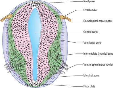
- In the fetus, multi-potent stem cells divide in the ventricular zone, generating neurons and radial glia. These glia act as scaffolding for neuroblast migration and inside out development of the cerebral cortex.
- These stem cells disappear by birth (likely differentiating into astrocytes). Some stem cells are left in specialised locations (e.g. hippocampus, olfactory bulb)
PNS patterning
Neural crest cells migrate from the dorsal neural tube to form most of the PNS (enteric, autonomic & sensory ganglia; schwann cells; adrenal medulla). They also form other structures such as head bones and mesynchyme.
Sensory PNS in the head arise from cranial neurogenic placodes, patches of ectodermal thickenings in the embryonic head.
Nervous system wiring
Axon growth and guidance
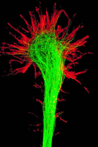
Connections in the nervous system are wired up by axon (and to a lesser extent dendrite) growth and guidance. At the tip of the each neurite (preliminary axon/dendrite), a growth cone leads the way to the correct destination, crawling like an ameoba via myosin and actin interactions. The growth cones are guided by several adhesion and signalling molecules:
- Diffusible
- Attractant: NGF, netrin (classed into nourishing ‘neurotrophins and attractant ‘neurotropins’
- Repellant: Semaphorins, Slit
- Contact
- Attractant: cell adhesion molecules (CAM) on growth cones adhere to ECM proteins (e.g. fibronectin), other CAM. Intracellularly, CAM connects to the cytoskeleton and cytoplasmic signal transducers
- Repellant: Sematophorins, ephrins, some proteoglycans
This chemical nature of axon growth and guidance was originally theorised as the chemoaffinity hypothesis. It is especially important in forming topographic maps (where a sensory or motor system is projected onto the CNS). One example is the retino-tectal projection where the each point on the retina can be mapped out on the tectum. When the optic nerve is cut in animals, the corresponding axons regrow to their original tectal positions by chemical gradients although final synapses are more variable and branch from these axons.
Neuronal selection
Once axons reach their destination, axons form synapses via synapse-specific adhesion molecules and chemical markers. Yet synapses are redundant and inefficient. These need to be refined and trimmed by activity-dependant mechanisms, involving many neuron deaths. This occurs via two main methods:
- via neurotrophic factors (mainly PNS): The number of neurons depend on supply of neurotrophic factors needed for growth. This is demonstrated by animal studies transplanting a limb bud resulting in more motor neurons. Here, secreted NGF binds to TrkA receptors on growth cone, sent back to cell body (by retrograde transport) to prevent apoptosis.
- via correlated activity (mainly CNS)
- Select for correlated activity: If presynaptic stimulation commonly result in postsynaptic firing, this synapse becomes strengthened (Hebb’s rule: “fire together, wire together”).
- Select against uncorrelated activity: Presynaptic stimulation does not commonly induce postsynaptic firing (i.e. gets cancelled out, “out-of sync, break the link”)
