
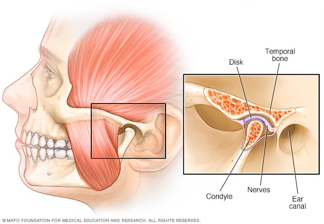
Each temporomandibular joint (TMJ) is a condyloid joint between the head of the mandible and the articular tubercle on the inferior surface of the temporal bone. As an atypical synovial joint, an intra-articular fibrocartilaginous disc divides the joint into upper and lower cavities and its articular surfaces are covered by fibrocartilage instead of hyaline cartilage.
Mechanics of mandibular movement
Movements of the TMJ normally result from some combination of the two components:
- Gliding in the upper compartment – the mandibular condyle and the intra-articular fibrocartilaginous disc move together over the articular surface.
- Hinge movements in the lower compartment, between the condyle of the mandible and the inferior surface of the fibrocartilaginous disc.
When opening the mandible fully, tightening of the temperomandibular ligament between the zygomatic arch and mandibular condyles shifts the axis of rotational movement shifts from the condyles initially to the lingula of the mandibular foramen where the sphenomandibular ligament attaches.
CLINICAL CORNER
Anterior dislocation of the temporomandibular joint may occur when the mouth is opened too wide (e.g. an excessive yawn or a punch). Under a light anaesthetic, the mandible is pressed downwards and backwards to lock it back into place.
