

The masseter, temporalis as well as medial and lateral pterygoid muscles primarily mediate mastication. They are all innervated by the mandibular nerve (CN Vc).
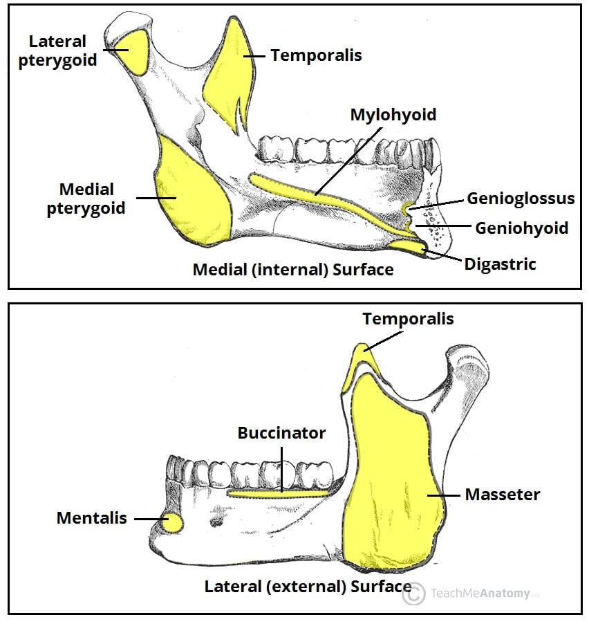
The masseter, temporalis and medial pterygoid elevate the mandible while lateral pterygoid is the only muscle that depresses the mandible:
- Masseter arises from the zygomatic arch and inserts into the lateral aspect of the ramus and angle of the mandible.
- Temporalis arises from the inferior temporal line and temporal fossa to insert into the coronoid process and anterior border of the mandibular ramus. It has a secondary action with posterior fibres retracting the mandible.
- Medial pterygoid arises from the pterygoid fossa and medial surface of the lateral pterygoid plate of the sphenoid to insert into the medial surface of the mandibular ramus. It can produce lateral movement when chewing (for molar grinding).
- Lateral pterygoid has two heads and is the only one that opens the mouth by protruding and depressing the mandible. Its superior head arises from the infratemporal fossa of the sphenoid and attaches to the articular capsule and disc of the TMJ (helping stabilise the joint). The inferior head arises from the lateral surface of the lateral pterygoid plate, inserting posteriorly to the pterygoid fovea (pit) on the neck of the mandible.
