

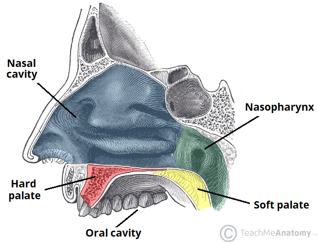
The palate forms the roof of the oral cavity (separating it from the nasopharynx) and consists of the hard palate anteriorly and the soft palate posteriorly.
Hard palate
The hard palate is covered with mucous membrane and formed by the palatine process of the maxilla and the horizontal plate of the palatine bone. Posteriorly, it is continuous with the soft palate.
Soft palate


Five muscles attach to and change the shape of the soft palate. These are the palatoglossus, palatopharyngeus, tensor (veli) palati, levator (veli) palati and musculus uvulae. Excluding tensor palati which is innervated by the mandibular nerve (CN Vc), these muscles are all innervated via the pharyngeal plexus by the pharyngeal branch of the vagus nerve (CN X).
- Palatoglossus and palatopharyngeus (which respectively insert into the tongue and the upper border of the thyroid cartilage) primarily raise either the tongue or oropharynx towards the palate to control the opening of the oropharynx.
- Tensor palati arises from the base of the medial pterygoid plate and Eustacean tube, its tendon coursing around the plate’s hamulus (an inferior hook-like process) to insert onto the sides of and tense the soft palate.
- Levator palati, arising from the inferior surface of the petrous temporal bone and the eustachian tube, elevates the soft palate to close the nasopharynx.
- Musculus uvulae shortens and broadens the uvula, changing the contour of the soft palate and also helps nasopharynx closure.
