

The middle ear (typmanic cavity) is a hollowed-out space in the petrous temporal bone. Primarily, its function is to transmit vibrations of the tympanic membrane via the ossicles to the inner ear. It is supplied by branches of the internal carotid and maxillary arteries, whilst innervated by the glossopharyngeal nerve through the tympanic plexus.
Boundaries of the middle ear
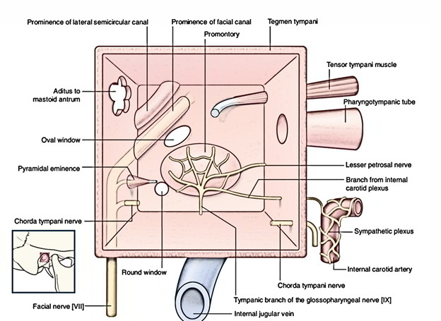
Its roof is a plate of bone separating the middle ear from the middle cranial fossa. Similarly, its floor is a thin plate of bone separating the cavity from the jugular fossa, which houses the start of the internal jugular vein and carotid canal.
The lateral wall is formed by the tympanic membrane. The chorda tympani passes posteriorly to anteriorly across the membrane and between the malleus and incus. Meanwhile, the medial wall consists primarily of the oval window which leads to the internal ear and the promontory (a bulge produced by the first turn of the cochlea). There is also the facial nerve which runs posteroinferiorly in the bony facial canal.
Posteriorly, the tympanic cavity communicates via the aditus with the mastoid antrum, a hollow air-filled cavity. The antrum is small at birth but enlarges as the mastoid process enlarges. It is inferior to the temporal lobe of the brain and separated by a thin plate of bone from the posterior cranial fossa.
From the anterior wall, the auditory (Eustachian) tube connects the middle ear with the nasopharynx, allowing air to enter or escape in order to equalise pressure between the cavity and the external environment. It has a medial cartilaginous part and a lateral bony part.
CLINICAL CORNER
Infection can pass from the nasopharynx to the middle ear via the auditory tube, resulting in middle ear infection or otitis media (common in children). Untrated, the tympanic membrane may rupture, a mastoid abscess could occur leading to meningitis and cerebral abscesses (as thin bone separated it from the cerebellum and temporal lobe).
Ossicles

The middle contains the three auditory ossicles – the malleus, incus, and stapes – which articulate with each other to transmit vibrations of the tympanic membrane to the inner ear.
The malleus has a lateral process and handle which are attached to the tympanic membrane, and a rounded superior head which articulates with the body of the incus. The incus articulates via its long process with the stapes. The base (footplate) of the stapes sits in the oval window (fenestra vestibuli). Movement of the base will vibrate the fluid in the internal ear, stimulating auditory receptors.
Muscles of the middle ear
The stapedius (innervated by nerve to stapedius from CN VII) and the tensor tympani (innervated by medial pterygoid nerve from CN V3) attach onto the ossicles to dampen loud sounds. The stapedius arises from the pyramidal prominence from the posterior wall of the middle ear and inserts into the neck of the stapes. The tensor tympani arises from the auditory tube and inserts into the handle of the malleus.
