
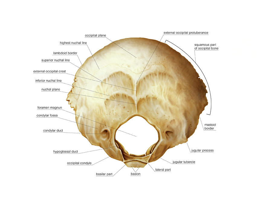
The occipital bone lies centrally in the posterior cranial fossa and is divided into squamous (flat) and basal parts. It contains the hypoglossal canals (for CN XII) and foramen magnum (for brain stem and vertebral arteries). Other landmarks include the occipital condyles (which articulate with the first vertebra atlas), occipital protuberance and nuchal lines (the superior of which attaches to the trapezius muscle).
