
The brain is supplied by the internal carotid and vertebral arteries which anastomose to form the circle of Willis at the skull base. These are not the sole intracranial arteries as branches of the external carotid artery are a major supplier to the meninges. Intracranial venous drainage does not follow the course of these arteries and instead run in the dural venous sinuses.
Internal carotid arteries

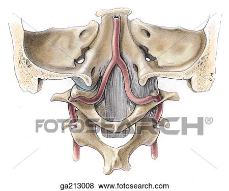
Each common carotid artery travels up the carotid sheaths before bifurcating into the internal and external carotid arteries at the level of the thyroid cartilage’s upper border. The internal carotid artery remains within the sheath to enter the cranial vault through the carotid canal, bending at right angles anteromedially to exit superior to foramen lacerum. It then turns superiorly and anteriorly into the cavernous sinus where it travels centrally alongside the abducens nerve (CN VI). Turning upwards to exit the sinus into the subarachnoid space, it gives off the ophthalmic artery before bending backwards and laterally to the lateral edge of the clinoid process. There, it turns superiorly towards the brain giving of anterior and middle cerebral branches.
Vertebral arteries
The vertebral arteries arise from the subclavian arteries and travel up the transverse foramina of cervical vertebrae, bending posteriorly and grooving the atlas (C1) before entering the cranial cavity through the foramen magnum.

Travelling anteriorly to the brain stem, each vessel gives off a small meningeal branch, branches to the medulla, the posterior inferior cerebellar artery, a posterior spinal artery and a branch to the anterior spinal artery. The two vertebral arteries then join to form the basilar artery which grooves the pons ventrally while giving off theanterior inferior cerebellar, labyrinthine, pontine and superior cerebellar arteries before dividing into the posterior cerebral arteries that supply the occipital lobes and form part of the circle of Willis.
CLINICAL CORNER
Vertebrobasilar insufficiency/ischaemia is caused by decreased blood flow to the posterior brain (e.g. a head turn which occludes the ipsilateral vertebral artery). Symptoms can include vertigo and blurred/double vision.
Circle of Willis
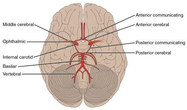
The circle of Willis is the anastomoses between the vertebral and internal carotid arteries around the optic chiasm and pituitary stalk. The anterior communicating artery connects the anterior cerebral arteries while the posterior communicating arteries connects each posterior cerebral artery with their ipsilateral internal carotid artery.
CLINICAL CORNER

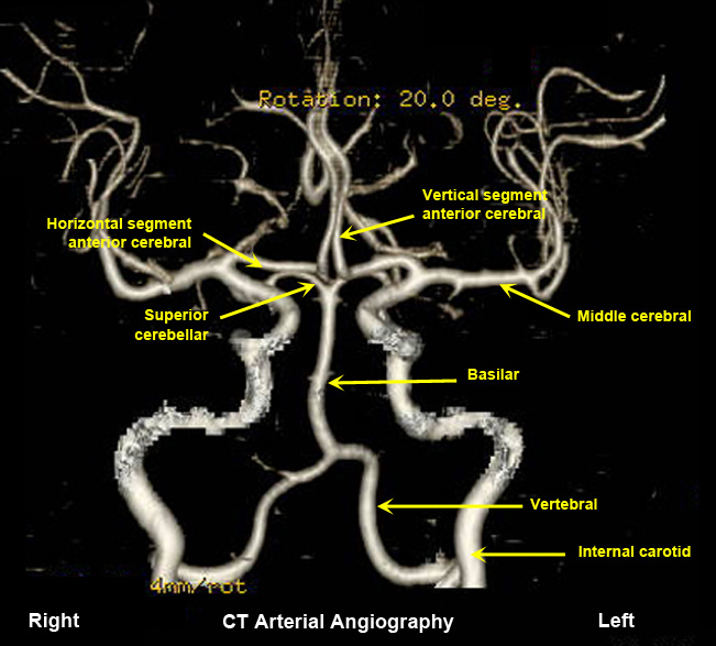
Cerebral angiography is used to examine arterial blood flow to the cranium.
